Acute L5 Intersegmental Subluxation
Acute lower back problems due to lumbar subluxations are common in our practices. They are rarely written up in spite of their importance, challenges, and frequency. It’s important to publish them, particularly for novice chiropractors as many chiropractic colleges try to discourage them from adjusting acute back pain patients. Dr. Vance presents a case of acute L5 intersegmental subluxation with a radiographically-viewed difference.
A 23-year-old male presented to our office on July 2, 2007, with acute lower back pain. He stated that he was racing a 4-wheeler two days previous and was thrown off. He had no pain after the accident and raced again the next day. The following morning, his pain was so severe that he couldn’t get out of bed.*
Upon examination, static palpation revealed extreme tenderness of L5. Due to the intensity of the pain, movement was limited on motion palpation with exacerbation upon right lateral bending and extension of the lumbar spine. An instrument reading was evident at L5. Achilles and patellar deep tendon reflexes were intact.
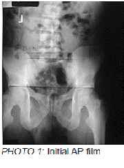
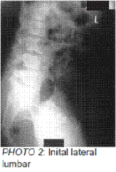
AP and lateral Lumbar x-rays showed a L5 PRS-M [photos #1 & #2]. The lateral x-ray demonstrated an intersegmental subluxation of L5. Noted was a wide disc (vertical height – diverging endplate lines at the posterior) at the posterior aspect of L4 and a loss of the lumbar lordosis. [photo #2] Uncharacteristic of these cases, L5 showed a wide disc at the posterior aspect. Typically in acute L5 subluxation, there is narrowing of the L5 disc at the posterior aspect, i.e., posteriorly converging endplate lines on the lateral film. There was also a noticeable retrolisthesis of L5.
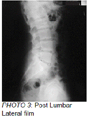
The side posture is recommended in acute lumbar subluxations as prone adjustments may exacerbate the pain. An adjustment to L5 was given on the pelvic bench with a pull move. The patient was instructed to use a cold pack for 20 minutes and to repeat after two hours if necessary. The patient returned the next day and reported slight improvement. The same adjustment procedure was repeated. After the adjustment, the patient stated that he felt much better. The patient missed his next appointment. He returned to our office on July 25, 2007, and stated that he felt great since his last adjustment. A lateral lumbar x-ray was taken to confirm correction. [photo #3] The lateral lumbar x-ray revealed a normal lumbar curve and better disc/vertebral placement.
In acute lower back cases such as this example, the patients are usually in extreme pain. Any pressure on L4 will exacerbate their condition. In this case on the lateral film, L4 disc space was open at the posterior and the lumbar curve was lost as well. Often there is extreme posterior-inferior misalignment of L5. In this presentation, the L5 disc space was wider at the posterior. This may be due to splinting of the lumbar musculature that results in loss of the lumbar curve or a D1 swollen disc that primarily affects the posterior disc.
This case is an excellent example of the effects of following the Gonstead protocol and the effectiveness of the Gonstead System.
[*Ed: an example of DOMS: “delayed-onset muscle soreness” (1-3 days delayed onset) in addition to acute joint injury which itself may take a day or two to manifest.]


