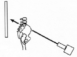
Angulated Lumbosacral View
Having trouble visualizing the sacroiliac joints on your AP film? Does one or both of the sacroiliac joints appear hazy or narrow on your AP film? Do you have questions about the shape of the L5 vertebral body? Consider taking an angulated lumbosacral view. Typically, if the sacroiliac joints are in doubt, one takes left and right obliques. You can reduce it to one view taken straight-on with the angulated lumbosacral view. If taken properly, one gets a great view through both sacroiliac joints and a great plane line view of the vertebral body of L5.
PROCEDURE
Measure the lumbosacral angle on the lateral x-ray. That becomes the angle of tube tilt. If you happen to be taking the view at the same time as the AP and lateral film, and the patient appears to have a normal sacral base angle, one can usually assume the angle is between 35°-40°. This view can be taken PA or AP. The PA view is taken with the tube pointed downwards. The AP view is taken with the tube pointed upwards.
Central ray is through L5.
MAS is approximately 1.5x the standard AP view. Kv is the same.
You will probably have to move the tube forward in order to get the proper angulation. You will have to adjust the exposure setting appropriately to compensate for changes in the FFD. I do it AP. Because my tube does not drop down too far, I have the patient stand on a foot stool, and I move the tube (FFD) up to 48” or so.
It can be taken on a 10×12 sheet of film, although you may risk losing the entirety of the sacroiliac joints. If in doubt, use a sheet of 14×17 film. Of course, collimate down.

Central ray goes through the plane line of the disc of the L5 disc. Raise the cassette so that the central ray is at the center of the film.
