Enteric Nervous System: A Review
Most of us have learned that there are two primary parts of the autonomic nervous system: the sympathetic and parasympathetic nervous systems. Neuroscientists have been describing a third part of the autonomic nervous system, the Enteric Nervous System (ENS). It functions relatively autonomously, i.e., it can function with minimal extrinsic control. Gershon calls the enteric nervous system: the “second brain.” (7) There may be some truth in the expression: “thinking with one’s stomach.” This is a brief “taste” of the enteric nervous system.
Structure of the Enteric Nervous System
Intrinsic Nerve Supply
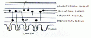
The enteric nervous system begins at the middle third of the esophagus and extends a distance of ten meters to the anorectal junction. (3) Some 100 million intrinsic neurons are embedded in the wall of the gut, an equal number to that in the spinal cord. (3,5,6) About 2,500 nerve cells are located in each millimeter of length of gut. (11) The enteric nervous system works independently of external input. The extrinsic nerve supply modifies its actions. (11) The enteric nervous system is composed of the myenteric or Auerbach’s plexus and the submucosal plexus of Meissner. (7) The myenteric plexus is a single complex found between longitudinal and circular smooth muscle layers of the muscularis externa and is involved with control of motility and the secretion of digestive enzymes. (3,7,11) The submucosal plexus is layered into inner, outer, and sometimes intermediate plexuses and has both sensory and motor neurons – the former communicates with the myenteric plexus, and the latter stimulates secretion from the epithelial crypt cells that release digestive juices into the gastrointestinal lumen. (7,11) Surprisingly, there are no nerve receptors in the lumen itself, and stimulus occurs transepithelially from various cells in the luminal and villi walls, such as, mucosal mast and enterochromaffin cells. (8)
The interstitial cells of Cajal are located between the neuronal terminals and the smooth muscle cells and are thought, by some, to be intermediary connections between the two. It has both inhibitory and excitatory transmitter receptors. (11) The Cajal cells are thought be the gastrointestinal equivalent of a “pacemaker” as they control peristalsis. (7) Like the “pacemaker” in the heart, the Cajal cells can propagate peristaltic waves without extrinsic neural control. (3)
Extrinsic Nerve Supply
The vagus nerve is a pathway for impulses between the gut and the brain. About 80%-90% of the vagus nerve functions as a visceral afferent nerve. (3,9FitzGerald 1985, Grundy 2006) The visceral afferent supply goes to the nucleus solitarius in the medulla oblongata. The visceral efferent supply comes from the dorsal nucleus of the vagus. (3) Gershon explains that the vagus nerve commands the ENS to carry out its specific tasks but it cannot tell the ENS how to carry out the tasks. (7)
The vagus nerve sends information to the central nervous system on the contents of the lumen and motor activity in the gut. (9) Afferent vagal impulses are involved in satiety, vomit reflex, and abdominal pain, among other brain-interpreted functions, in the presence of food in the lumen or the presence of toxins or pathogens. Vagal efferent/motor nerves have been found to innervate neurons in the myenteric plexus that release serotonin or vasoactive intestinal peptide. (7)
The vagus nerve contributes vagal motor neurons to the striated muscles of the esophagus, sympathetic noradrenergic neurons to the gut muscles, and noradrenergic vasoconstrictor neurons that innervate arteries within the GI tract walls. (4)
Sympathetic preganglionic fibers that innervate the stomach and on down to the splenic flexure of the colon come from spinal cord segments T5 to T12 through the thoracic splanchnic nerves. These terminate in the abdominal prearotic splanchnic ganglia. Postganglionic fibers then travel with the gastrointestinal arteries. (3) The lumbar sympathetic chain ganglia supply preganglionic fibers to the splanchnic ganglia which supply the descending and sigmoid colon and the rectum. (3)
The sympathetic spinal nerve supply to the gut processes noxious stimuli that results from tissue injury, ischemia, and inflammation. (9) In response to damage of the mucosa, spinal afferent neurons can initiate a local response. It causes neurogenic inflammation by increasing blood flow and vascular permeability, as well as, increases mucus and bicarbonate secretions and reduces acid secretion. (10)

Neurotransmitters
As we now know, the primary neurotransmitters that are associated with the sympathetic nervous system are epinephrine and nor-epinephrine. In the parasympathetic nervous system, it is acetylcholine. Every type of neurotransmitters and transmitter-type chemicals have been found in the enteric system, including substance P, adenosine triphosphate (ATP), cholecystokinin, and enkephalins. (3,8)
Gershon has found that over 95% of the 5-hydroyxtryptamine (5-HT or serotonin) in the body is in the enterochromaffin cells of the gastrointestinal epithelium with a small amount in the enteric interneurons. (7,8) Serotonin is released when the peristaltic reflex is engaged and appears to stimulate the reflex. (7) The enterochromaffin cells also “tastes” the luminal contents. (9) 5-HT is released in the presence of toxins and causes the dilution and elimination of toxins in the gut via vomiting or diarrhea and prevents further ingestion with nausea. (9) In addition, sugars in the lumina engage in the release of 5-HT and must be reduced to monosaccharides for absorption. (9,13) The serotonin is released spontaneously by the enterochromaffin cells into the lamina propria or as a result of increased intraluminal pressure or exposure to other neurotransmitters or chemical, among other things. There are at least 15 subtypes of serotonin receptor in the propria. (7) When the serotonergic receptors of the IPAN are stimulated, these IPANs release acetylcholine and calcitonin gene-related peptide which stimulates myenteric neurons, which in turn, stimulate either serotonergic interneurons or the enteric musculature. The serotonin released also stimulates vagal extrinsic afferent nerves that pass on to the central nervous system. (8.9) The large quantity of serotonin released by the enterochromaffin cells may be due to the distance from these cells to the target nerve receptors, as well as, the high turnover rate of epithelial cells in the gut. (8) Without an extracellular enzyme to catabolize serotonin, enterocytes reuptake it. (8) Those receiving radiation treatments for cancer usually get nausea and vomiting when serotonin leaks out of the enterochromaffin cells and activates 5-HT3 receptors. (7) Many ingest psychoactive drugs that affect serotonin, e.g., SSRIs, have gut problems.
Cholecystokinin (CCK) is a hormone and neuropeptide that is involved in the regulation of the digestive process. It is secreted in the proximal small intestines in response to food – particularly fat and protein – leaving the stomach. (12) Among its many activities, it has been found to stimulate gallbladder contraction and pancreatic enzyme secretion, induce satiety and reduce food intake, inhibit gastric emptying and gastric acid secretion, and stimulate intestinal peristalsis. (12) CCK is involved in signaling the process to break down long chain lipids to an absorbable form. (13) In the gut, the CCK-expressing neurons are found in both the myeteric and submucosal plexuses, pancreas, vagus nerve, among many other neurons. (12) In the gut, it is produced primarily by the endocrine cells of the small intestine. In the nervous system, the majority of CCK-expressing neurons are in the brain, in particular, the cerebral cortex. (12) Those found in the hypothalamus are thought to be involved in the sensation of satiety. (12) It is a important factor in the gastric emptying of solid and liquid meal from the stomach. (1)
How It All Works (Probably)

One of the most enduring images of digestion is a boa constrictor that has consumed a medium-sized mammal. The image is a long tube with a huge bulge in the middle. How does this huge object go from a whole critter to bones and other matter upon exit and how does it progress from mouth to anus. As you might well understand, some very complex processes are at work, and the nervous system is controlling the process. I am using the snake for visualization purposes only as its gut is anatomically and physiologically different from a human’s. How the enteric nervous system functions is not fully understood. The description below is what current science believes to occur.
Once food enters the mouth and mastication begins, saliva is produced and begins the digestive process. Obviously, there are afferent and sensory inputs that travels to the central nervous system (the mouth is outside of the scope of this paper). The input to the central nervous system does prepare the rest of the GI tract for the oncoming food. It is said that the better the mastication of food in the mouth, the better the digestive process through the rest of the gut, both through better initial breakdown of food and initiating signals the central nervous system to begin digestion.
Several important processes must occur during digestion: 1) propelling the food from mouth to anus; 2) detect the chemical constituents of the food and detect noxious chemicals; 3) secrete digestive juices; 4) mash or “chop” the food and mix it with digestive juices and absorb nutrients into the circulatory system; 5) eliminate waste products; and 6) control the vascular supply to the gut.
How is Food Propelled in the Right Direction?
To propel the food bolus, there is a coordinated process of muscle contraction and relaxation. The muscles are annular rather than longitudinally oriented, i.e., when they contract, they squeeze the lumen. Whatever is in front of the contracting muscle is propelled forward – there must be contraction on the oral side and relaxation at the site of distention and on the anal side. This circuit requires sensory and afferent nerves, interneurons, and motor neurons. Most of the intrinsic primary afferent neurons (IPAN) are in the myenteric plexus. Most of these afferent neurons appear to be affected by contraction or tension of the gut wall muscles rather than distention by the food bolus. (11) There is a bit of the “chicken and the egg” scenario as the muscles require neurological control. Excitatory stimuli of these adjacent neurons and also the saturation by serotonin and the firing of excitatory interneurons are probably factors. In the latter situation, there is a complex interconnection of IPANs and three levels of interneurons that ultimately stimulate motor neurons that contract gut muscles. (11) From various studies, neurons that control the mixing process affect 10-20 mm of gut while those controlling peristalsis control a longer region of gut. (11)
How is that poor critter being pushed through the gut of the snake. At the point of distention, gut circular muscles are relaxed, therefore the IPANs are partially inactive. At the point of distention, there is a localized accommodation reflex whereby inhibitory motor neurons are activated and relax muscles. (11) Ascending excitatory interneurons initiate a reflex back to motor neuron behind the bolus that initiates contraction. IPANs behind the bolus are also activated when sensing muscle tension and further stimulate motor neurons. Forward of the bolus, descending inhibitory interneurons cause a reflex action which along with muscle relaxation partially inactivates IPANs ahead of the bolus. With muscle relaxation ahead of the bolus, there is inactivation of ascending excitatory reflexes. (11) The muscle contractions behind the distention and relaxation at the point of distention and forward of it allows the bolus to proceed forward on this complex dis-assembly line.
Breaking Down of Food
There are three primary processes involved in extracting useful nutrients from food: the release of digestive juices, the processes of “chopping” food matter, and mixing of digestive juices with the bolus for the extraction of nutrients. Control of these processes are functions of the enteric nervous system. As noted above, the neurons controlling the mixing process affect a shorter length of gut than those involved in peristalsis. This is probably due to the fact that a greater degree of muscular contraction is involved.
Secretomotor stimuli come from mechanical or chemical changes in the lumen and motility changes in the small intestines. (11) The neural mechanism involves nerve endings in the mucosa from IPANs and the mysenteric and submucosal plexuses. (11) These reflex back via secretomotor neurons whose cell bodies are in the submucosal plexus. Vasomotor activity to provide vascular support to the activities of the of the GI tract is not well known. Vasodilator reflexes are stimulated by mechanical or chemical changes in the mucosa. (11) Secretomotor and vasomotor functions appear to come from the same neurons. (11)
Other sensations utilize various routes to the central nervous system. The following obviously require external nerve pathways to the central nervous system. The sensation of well-being or pleasure after a meal is undoubtedly related to multiple neural and hormonal stimulations with smell, taste, and gastric distention among the major factors. Distention of the rectal area likely causes the urge to defecate with the addition of noxious stimulation if the distention persists. Nausea and the urge to vomit have a complex of neural afferent and efferent pathways. Visceral GI tract pain appears to be related to receptors that, at lower levels of stimulation, cause reflex actions. At higher levels of stimulation, they cause a noxious sensation and the activation of nociceptors that solely fire with noxious input. (2)
When one considers the variety and complexity of activities in the GI tract – the process of digestion and extraction of nutrients, hunger, satiety, toxins or pathogens, vomiting and diarrhea mechanisms, nausea sensation, and changes by or to behavior, exact coordination and communications by the various parts of the nervous system is obviously vital. The nervous system must function optimally for proper GI tract function.
The space-limited brevity of this article barely touches the topic but hopefully whets your appetite for further study yourself`. A more comprehensive overview article is being prepared for the GCSS web site. More on the autonomic nervous system at Meeting of the Minds IV.
References
1 Borovicka J, Kreiss C, Asal K, et al. Role of Cholecystokinin as a Regulator of Solid and Liquid Gastric Emptying in Humans. American Journal of Physiology September 1996; 271(3, Pt.1):G448-G453.
2 Cervero F. Neurophysiology of Gastrointestinal Pain. Baillière’s Clinical Gastroenterology January 1988; 2(1):183-199.
3 FitzGerald MJT. Neuroanatomy Basic & Applied. Philadelphia: Baillière Tindall. 1985. pp.266-275.
4 Furness JB. Types of Neurons in the Enteric Nervous System. Journal of the Autonomic Nervous System 2000; 81:87-96.
5 Furness JB, Costa M. Types of Nerves in the Enteric Nervous System. Neuroscience January 1980; 5(1):1-20.
6 Furness JB, Kunze WAA, Bertrand PP, et al. Intrinsic Primary Afferent Neurons of the Intestine. Progress in Neurobiology 1998; 54:1-18.
7 Gershon MD. The Enteric Nervous System: A Second Brain. Hospital Practice 15 July 1999; 34(7):31-32, 35-38, 41-42, 46-47, 51-52.
8 Gershon MD. Nerves, Reflexes, and the Enteric Nervous System: Pathogenesis of the Irritable Bowel Syndrome. Journal of Clinical Gastroenterology May/June 2005; 39(Suppl. 3):S184-S193.
9 Grundy D. Signaling the State of the Digestive Tract. Autonomic Neuroscience: Basic and Clinical April 2006; 125(1-2):76-80.
10 Holzer P. Efferent-like Roles of Afferent Neurons in the Gut: Blood Flow Regulation and Tissue Protection. Autonomic Neuroscience: Basic and Clinical April 2006: 125(102):70-75.
11 Kunze WAA, Furness JB. The Enteric Nervous System and Regulation of Intestinal Motility. Annual Review of Physiology 1999; 61:117-142.
12 Liddle RA. Cholecystokinin Cells. Annual Review of Physiology 1997; 59:221-242.
13 Raybould HE, Glatzle J, Freeman SL, et al.
